
Inside Aging Blood Vessels: Scientists Map the Cellular Changes That Develop Over Time
Key Points
Researchers assembled a Human Aging Endothelial Cell Atlas using data from more than 375,000 cells, 290 samples, 12 human tissues and 34 existing studies.
The study examines how endothelial cells, which line the inside of blood vessels, change their gene activity with age.
Older individuals showed reduced activity of several genes involved in blood-vessel formation in cells lining veins.
Age-associated changes also involved genes linked to inflammation, cellular senescence and mechanical-force sensing.
The findings provide a research resource for investigating vascular aging, but they do not establish that the observed molecular changes directly cause blood-vessel deterioration.
advertisement
A New Look at How Blood Vessels Age
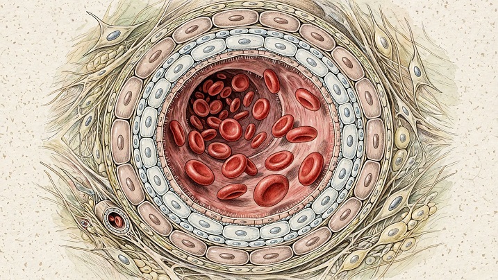
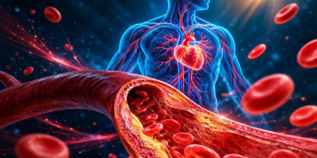
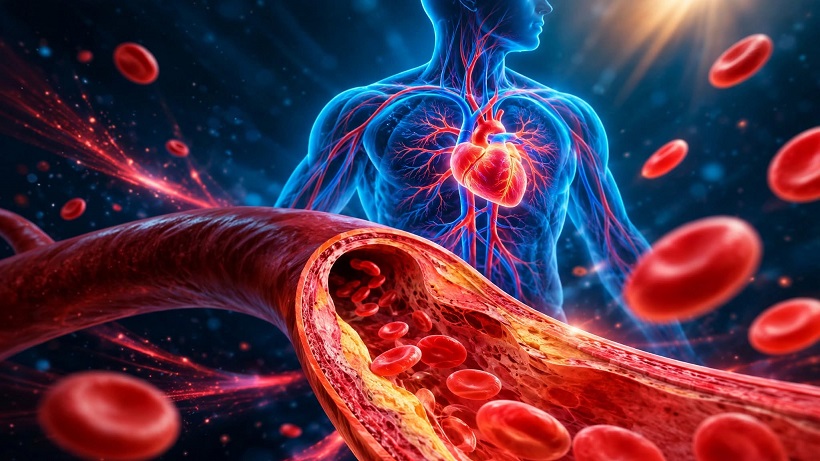
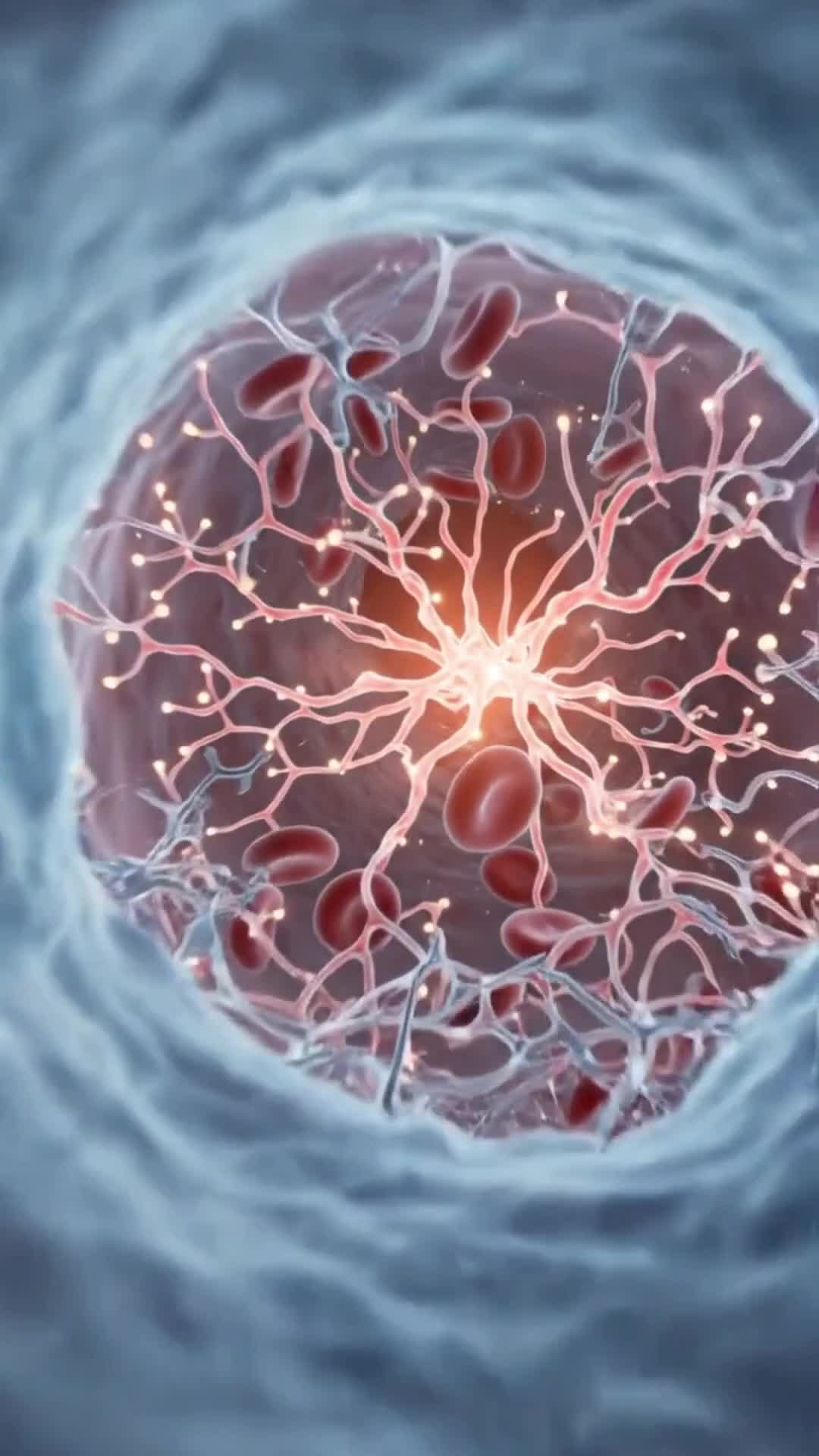
Blood vessels do more than transport blood around the body. Their inner surfaces are lined with specialized cells that help regulate blood flow, control the movement of substances into surrounding tissues and coordinate responses to injury. These cells, known as endothelial cells, also support the distinct functions of organs, including the protective barrier of the brain and the filtration processes of the kidneys.
As people grow older, the function of this finely coordinated system can deteriorate, increasing susceptibility to cardiovascular disease. Yet the molecular changes that occur inside endothelial cells during aging have not been fully understood. A study published in Nature Aging, led by researchers including first author Sarah Dobner and principal investigator Laura de Rooij at the CeMM Research Center for Molecular Medicine of the Austrian Academy of Sciences, investigates these changes across multiple human tissues.
Rather than treating blood vessels as a single structure, the researchers examined patterns of gene activity in individual endothelial cells. Their findings suggest that vascular aging involves several biological processes and differs across cell populations and tissues.
Mapping Aging Across More Than 375,000 Cells
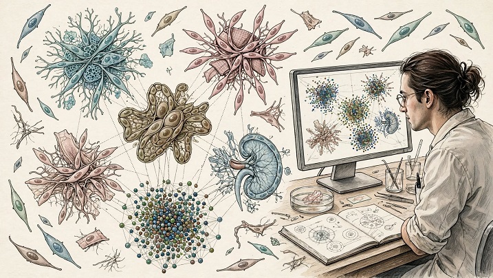
To investigate how endothelial cells change over time, the researchers combined data from 34 previously published studies, covering more than 375,000 cells from 290 samples across 12 human tissues. They concentrated on tissues classified as healthy or nondiseased to investigate molecular changes associated with aging rather than changes primarily attributable to diagnosed disease.
The resulting Human Aging Endothelial Cell Atlas provides a resource for examining how the activity of genes in blood-vessel-lining cells varies with age and across different parts of the body. This approach is important because endothelial cells do not perform identical functions everywhere. Cells serving different organs operate in distinct environments and must meet different physiological demands.
By integrating data from multiple studies, the researchers could investigate patterns that might be difficult to identify when examining a single organ or endothelial cell population in isolation. The atlas offers a broader view of vascular aging, although its findings remain dependent on the available samples and the quality and completeness of the underlying data.
Older Vein-Lining Cells Show Changes Linked to Vessel Formation
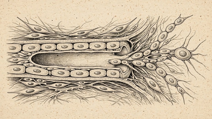
One notable finding involved endothelial cells lining veins. These cells can contribute to the formation of sprouts that develop into new blood vessels, a process involved in vascular renewal. In older individuals, the researchers observed reduced activity of several genes associated with blood-vessel formation in these cells.
The result raises the possibility that aging may reduce some of the molecular capabilities associated with building or renewing blood vessels. However, the distinction between a molecular signature and a demonstrated physical effect is important: the study did not directly measure the repair capacity of blood vessels. It therefore cannot establish that the observed gene-activity changes necessarily result in slower or less effective vascular repair.
The finding nevertheless identifies a specific area for further investigation. Understanding whether these changes contribute to declining vessel function, reflect other age-related processes or vary according to tissue could help researchers clarify how vascular aging develops.
Inflammation and Cellular Senescence Appear in the Aging Signature
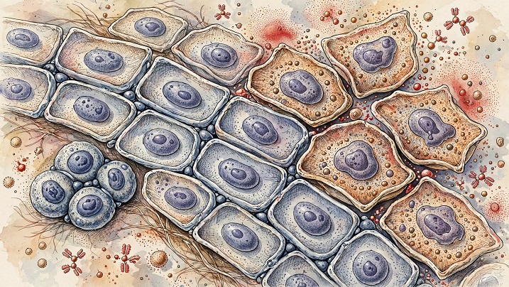
Across multiple tissues, the analysis also identified increased activity of genes associated with inflammation and cellular senescence. Cellular senescence describes a state in which cells stop dividing but can remain biologically active and influence their surroundings.
Senescence-related changes are relevant to aging research because they may affect the environment around a cell. However, identifying genes associated with senescence does not automatically establish that every cell showing those signals has entered a stable, permanent senescent state.
The researchers examined a group of genes associated with p21, a protein that can slow or stop cell division. Comparisons involving laboratory-grown endothelial cells and mouse models of accelerated aging supported a connection between these gene patterns and cellular senescence. Even so, the researchers noted that the genes involved can also participate in stress responses and blood-vessel growth.
This overlap makes interpretation difficult. A gene signature may indicate senescence-related activity, but it can also reflect other biological challenges experienced by aging cells. More precise methods will be needed to distinguish these possibilities before scientists can determine which processes might be suitable targets for future interventions.
How Endothelial Cells Respond to Their Physical Environment
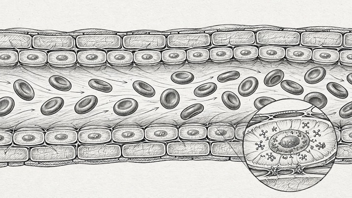
Blood-vessel-lining cells continually experience mechanical forces generated by flowing blood and by the material surrounding them. They sense these forces and translate them into biological signals that influence their behavior. The researchers found age-associated changes in genes involved in these responses, suggesting that the way endothelial cells interact with their physical environment may change over time.
Experiments using human skin provided an additional observation involving YAP, a protein that helps cells respond to mechanical forces. YAP was found more frequently inside the nuclei of endothelial cells from older donors. Because a cell's nucleus contains its genetic material, the location of a signaling protein can provide clues about changes in cellular activity.
The observation does not, however, establish whether increased nuclear YAP activity helps older endothelial cells adapt to their environment or contributes to deteriorating blood-vessel function. Both possibilities remain questions for further research. Determining the role of YAP will require experiments that move beyond observing age-related differences and directly test their biological consequences.
Why Vascular Aging Does Not Follow One Pattern
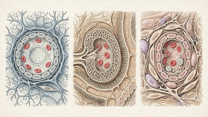
A central message of the study is that endothelial aging is not uniform across the vascular system. Different cell populations and tissues displayed different molecular patterns, reinforcing the importance of examining the blood-vessel lining in its specific biological context.
Some changes may be associated with declining function, while others could represent attempts by cells to cope with the physical and biological stresses of aging. These explanations are not interchangeable: a molecular change observed in older tissue does not necessarily mean the change is harmful. It may be a cause of dysfunction, a consequence of another process or part of an adaptive response.
The study's integrated approach provides a framework for exploring these alternatives. By comparing patterns across tissues and cell populations, researchers can identify questions for targeted experiments and investigate whether particular molecular changes are shared across the body or restricted to specific vascular environments.
What the Study Can—and Cannot—Tell Us
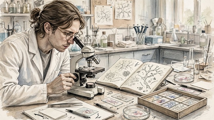
The findings offer a detailed resource for studying the molecular characteristics of aging endothelial cells, but several limitations matter when interpreting the results. The researchers compared people of different ages rather than following the same individuals over time. As a result, the analysis identifies age-associated differences but cannot directly trace how an individual's endothelial cells change throughout their life.
The available information about donors' lifestyles and medical histories was also incomplete, limiting the researchers' ability to account for every factor that might influence the observed patterns. Focusing on tissues classified as healthy or nondiseased helps investigate aging-related changes, but it does not eliminate every possible source of variation between samples.
These limitations mean that the results should not be interpreted as proof that a particular gene or protein causes vascular aging. Nor do they establish that changing YAP, p21-associated pathways or other observed signals would prevent cardiovascular disease or restore aging blood vessels. Those questions require additional research.
A Resource for Future Vascular-Aging Research
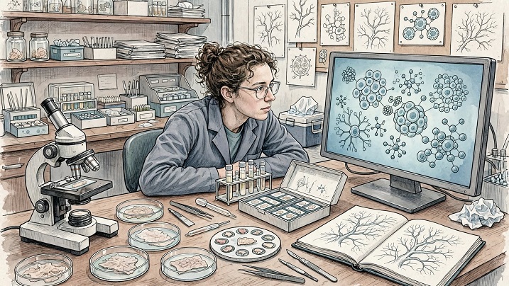
The Human Aging Endothelial Cell Atlas gives researchers a way to explore molecular changes across multiple tissues rather than treating blood vessels as a single, uniform system. An accompanying interactive portal described by the researchers is intended to help scientists investigate the atlas and identify directions for future experiments.
The next challenge is to establish which age-associated changes have functional consequences. Researchers will need to determine whether particular gene patterns contribute to declining vessel performance, represent protective adaptations or simply accompany other processes. They will also need to test whether findings observed in one tissue apply to endothelial cells elsewhere in the body.
The work does not present an established treatment for aging blood vessels. Instead, it offers a foundation for more focused investigations into how the cells lining blood vessels respond to aging and which mechanisms may be most important for maintaining vascular function over time.
Key Points Summary
Endothelial cells line blood vessels and help regulate circulation, tissue exchange and responses to injury.
Researchers combined 34 studies into an atlas covering more than 375,000 cells across 12 human tissues.
Older vein-lining endothelial cells showed reduced activity of several genes associated with blood-vessel formation.
Aging was associated with changes in inflammation-related, senescence-related and mechanical-response genes.
YAP was more frequently found inside endothelial cell nuclei in skin samples from older donors, but the consequences remain uncertain.
The study identifies molecular patterns associated with aging rather than proving that these changes directly cause vascular dysfunction.
The atlas is intended to support future research into the mechanisms of vascular aging.
What This Means
Why it matters: The study provides a broader picture of how blood-vessel-lining cells change with age and highlights differences across tissues that could be missed in narrower investigations.
Who may be affected: The findings are relevant to researchers studying aging, cardiovascular biology, endothelial function and the molecular processes associated with age-related disease. They do not establish an immediate change in clinical care.
What to watch next: Further experiments will need to determine which molecular changes contribute to impaired vascular function, which represent adaptive responses and whether any of these processes can be safely targeted in future research.
advertisement
Frequently Asked Questions (FAQ)
1. What are endothelial cells?
Endothelial cells form the thin inner lining of blood vessels. They help regulate blood flow, control the exchange of substances with surrounding tissues and coordinate responses to injury.
2. What did the new study investigate?
The researchers examined age-associated gene-activity patterns in endothelial cells across multiple human tissues, using data integrated from 34 existing studies.
3. How large was the research dataset?
The atlas incorporated data covering more than 375,000 cells from 290 samples across 12 human tissues.
4. What changes were observed in older vein-lining cells?
Older individuals showed reduced activity of several genes associated with blood-vessel formation. The study did not directly measure whether these cells were less capable of repairing blood vessels.
5. What is cellular senescence?
Cellular senescence is a state in which cells stop dividing but can remain active and influence their surrounding environment. The study identified gene patterns associated with senescence, but those patterns cannot reliably identify every permanently senescent cell.
6. What is YAP, and why is it relevant?
YAP is a protein involved in how cells respond to mechanical forces. In human skin samples, it was found more frequently inside the nuclei of endothelial cells from older donors. The study did not establish whether this difference is beneficial or harmful.
7. Does the study prove that aging causes blood vessels to lose their ability to repair themselves?
No. The study identified reduced activity of several vessel-formation-related genes in older vein-lining cells, but it did not directly test blood-vessel repair capacity or establish causation.
8. Could the findings lead to new treatments?
The atlas may help researchers identify mechanisms for future investigation. However, the study does not establish a treatment, demonstrate that targeting the identified pathways would improve vascular health or show that a new intervention is ready for clinical use.
Sources
Nature Aging — Primary research: Uncovering the transcriptional hallmarks of endothelial cell aging via integrated single-cell analysis. DOI: 10.1038/s43587-026-01247-3.
Medical Xpress — Supporting report: How our blood vessels age from within, October 27, 2026.
Additional Verified Sources
No additional sources used.
Thank you !