article image source: brown.edu (Link)
Glowing Neurons: How Bioluminescence is Revolutionizing Brain Imaging in Real Time
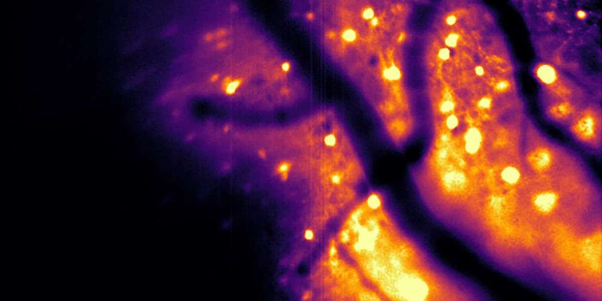
This video shows mouse cortical neurons flashing with bioluminescence while the mouse runs on a wheel. By Jeremy Murphy/Bioluminescence Hub.
Key Points Summary:
Brown University’s Bioluminescence Hub developed CaBLAM, a bioluminescent tool that allows real-time tracking of brain activity.
CaBLAM uses bioluminescence, which produces its own light, avoiding the risks of external light exposure like phototoxicity and photobleaching.
This method offers clearer, more precise images, enabling scientists to track individual neurons and subcellular processes for extended periods.
The tool could potentially be used to study other parts of the body and to control neural communication with light.
The Bioluminescence Hub's research has broad implications for understanding complex brain functions, learning processes, and developing new medical treatments.
advertisement
Introduction
Brain imaging has long been a critical tool for understanding the inner workings of the brain. From studying cognition to tracing the origins of neurological diseases, scientists rely on detailed images of brain activity. Traditional methods, such as fluorescence-based imaging, have made remarkable strides. However, these methods often come with significant limitations, such as photobleaching and potential harm to brain cells due to prolonged light exposure. But what if there was a way to visualize the brain’s activity without these drawbacks? The answer may lie in bioluminescence, a glowing solution that is transforming neuroscience.
Researchers at Brown University’s Carney Institute for Brain Science have pioneered the development of a new, innovative brain imaging tool that uses bioluminescent light to measure brain activity safely and effectively. This groundbreaking method promises to offer deeper insights into brain function, with fewer risks and greater accuracy than traditional methods. But how exactly does this work? Let’s dive into the science behind bioluminescence and its potential to revolutionize neuroscience.
This video, courtesy of collaborators with the Bioluminescence Hub,
captures HeLa cells in the lab that have been engineered to self-create light.
The Birth of the Bioluminescence Hub
In 2017, the Bioluminescence Hub was launched at Brown University with the goal of developing advanced neuroscience tools that would allow scientists to monitor and manipulate brain activity with light. Led by Christopher Moore, a professor of brain science, along with collaborators from Central Michigan University and the University of California, San Diego, the Hub set out to create methods that go beyond traditional fluorescence imaging.
One of their key innovations is the Ca2+ BioLuminescence Activity Monitor, or CaBLAM. This molecule, developed by Nathan Shaner at UC San Diego, is a shining example of how bioluminescence can be used in brain imaging. Unlike fluorescence, which requires external light sources like lasers, bioluminescence produces its own light. This eliminates many of the risks associated with other methods, such as phototoxicity and photobleaching, making it a safer and more efficient way to observe brain activity in living animals.
advertisement
How Does Bioluminescence Improve Brain Imaging?
The challenge with traditional fluorescence-based brain imaging lies in the need for external light sources. These light beams can damage cells and cause fluorescence molecules to lose their ability to emit light over time, a phenomenon known as photobleaching. In contrast, bioluminescence doesn’t require external light. Instead, it harnesses the natural ability of certain molecules to emit light when an enzyme breaks them down. This process is not only safer for the brain but also allows for clearer, more precise imaging.
According to Christopher Moore, bioluminescence has a distinct advantage over fluorescence because the brain naturally produces background light when exposed to external light. This “glow” can interfere with imaging signals, making it difficult to distinguish brain activity. However, with bioluminescence, engineered neurons produce their own light, standing out sharply against the dark background. This reduces interference and allows scientists to observe the brain’s activity in much greater detail, even at deeper levels of the brain.
CaBLAM: A Breakthrough in Real-Time Brain Activity Monitoring
The team’s development of CaBLAM marks a significant leap in neuroscience research. The CaBLAM tool enables scientists to track the activity of individual neurons and even monitor subcellular processes in real time. In their recent study published in Nature Methods, the team demonstrated the ability to record brain activity for up to five hours—a feat impossible with traditional fluorescence methods due to the time limitations imposed by fluorescence molecules.
CaBLAM's ability to capture continuous data over extended periods is crucial for studying complex behaviors and learning processes. For example, scientists can now study how neurons communicate over time, observing long-term changes in brain activity that were previously difficult to capture.
Moore emphasizes that this breakthrough is just the beginning. "We are now seeing single cells activated independently in a way that was never possible before. It’s like having a special, highly sensitive movie camera recording brain activity as it happens," he said. This advancement could be pivotal in understanding how the brain learns, remembers, and processes information.
advertisement
The Future of Bioluminescence in Neuroscience
The implications of bioluminescent brain imaging are far-reaching. Beyond monitoring brain activity, the technique could potentially be used to "rewire" the brain by using light to control neuronal activity. For example, one project within the Bioluminescence Hub explores how light could be used to control neuron-to-neuron communication. This could provide a non-invasive way to influence brain function, offering potential treatments for neurological conditions like epilepsy and Parkinson’s disease.
Moore envisions the application of this technology extending beyond the brain. “This tool could eventually allow us to monitor other parts of the body, enabling real-time tracking of how different organs interact with one another,” he said. The future of bioluminescence in brain and body research holds immense promise.
Conclusion: The Power of Team Science and Innovation
The work being done at Brown University’s Bioluminescence Hub exemplifies the power of collaborative scientific efforts. More than 34 researchers from across various institutions, including UC San Diego, Central Michigan University, and New York University, have contributed to the development of this transformative technology. Their work, supported by the National Institutes of Health (NIH), the National Science Foundation (NSF), and the Paul G. Allen Family Foundation, is pushing the boundaries of what is possible in neuroscience.
As we continue to explore the brain’s complexities, the ability to observe it in real time, with greater clarity and safety, will undoubtedly open new doors for research and medical treatments. The future of neuroscience is brighter than ever, and bioluminescence may be the key to unlocking even deeper insights into how our minds work.
FAQ
Q1: What is bioluminescence in brain imaging?
Bioluminescence in brain imaging refers to using light produced naturally by certain molecules to monitor brain activity. Unlike traditional methods that require external light sources, bioluminescence generates its own light, making it safer and more precise for studying brain function.
Q2: How does CaBLAM work?
CaBLAM is a bioluminescent molecule that allows researchers to observe brain activity in living organisms. It eliminates the need for external light, providing clearer and longer-lasting recordings of brain cell activity.
Q3: What are the benefits of using bioluminescence over traditional fluorescence imaging?
Bioluminescence doesn’t require intense external light, which reduces the risks of damaging cells or causing photobleaching. It also provides clearer images since it avoids interference from background light in brain tissue.
Q4: Can bioluminescence be used for other parts of the body?
While it is currently being used for brain imaging, bioluminescence has the potential to be applied to monitor other organs and systems in the body, providing new insights into how different parts of the body interact in real time.
Q5: How will bioluminescence impact medical research?
By offering a safer, more precise way to observe brain activity, bioluminescence could revolutionize the study of neurological diseases, cognitive functions, and potential treatments for conditions like epilepsy and Parkinson’s disease.
Sources:
Brown University: Is bioluminescence the key to safe, effective brain imaging?
https://www.brown.edu/news/2025-12-12/bioluminescence-brain-imaging-tool
Thank you !