Faster Imaging Reveals Hidden Brain Wiring
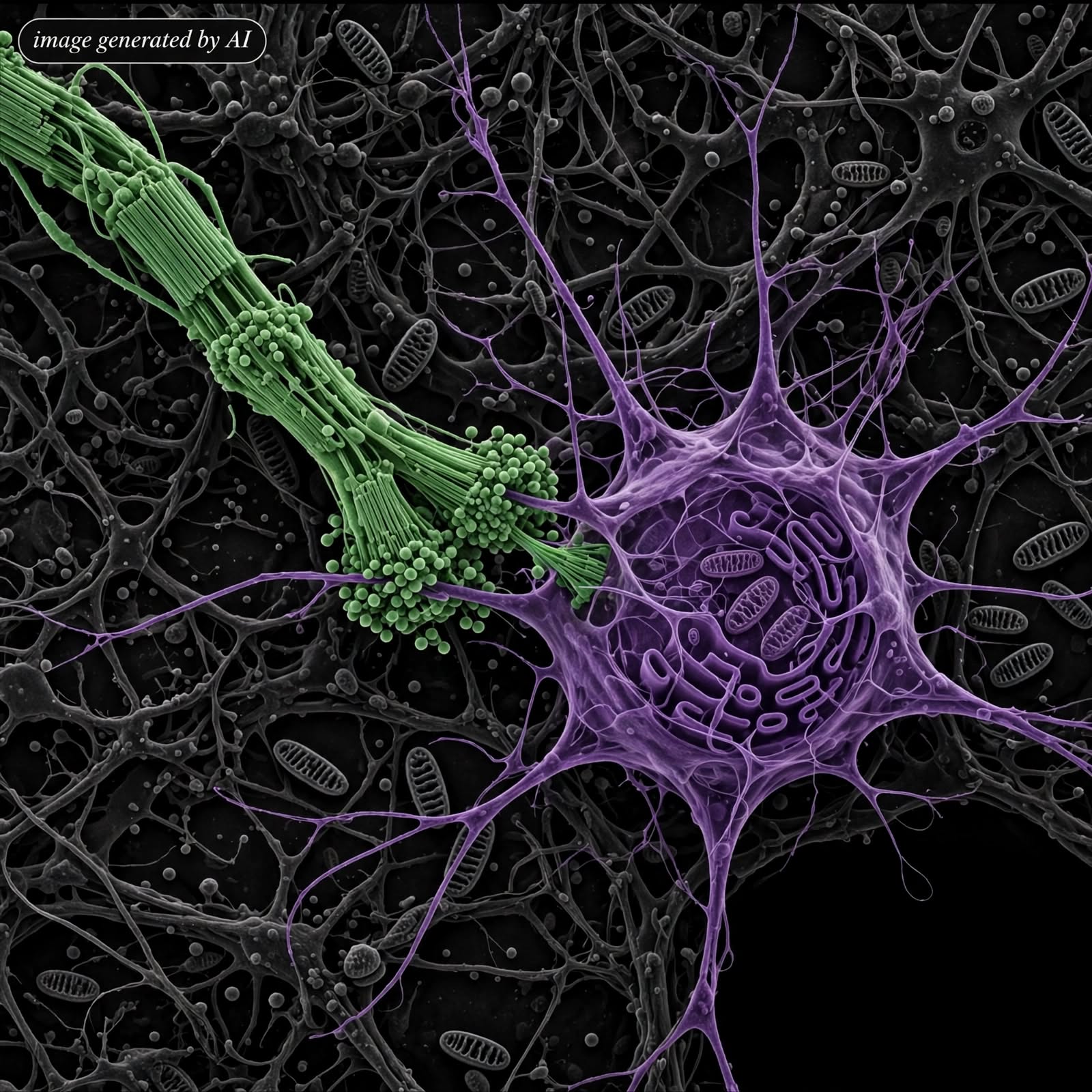
Researchers at Harvard Medical School and the University of California San Diego have sped up electron tomography to map volumes of brain tissue in detail. The new method uses about 50 times fewer images than standard tomography while working with tissue slices about 20 times thicker than usual. It still reaches 3D resolution near 15 nanometres and can reveal structures that many connectome maps miss. In mouse cerebellum tissue the team spotted electrical synapses that let signals pass almost instantly between neurons. They also saw tiny protrusions where neurons appeared to nibble material from neighbouring glial cells. Those exchanges may help shape how networks form and how cells share material. The team traced nerve fibres through a 3D block to the points where they meet other neurons. Scaling the approach to a whole mammalian brain will need automated sample handling but the result opens a clearer view of brain wiring.